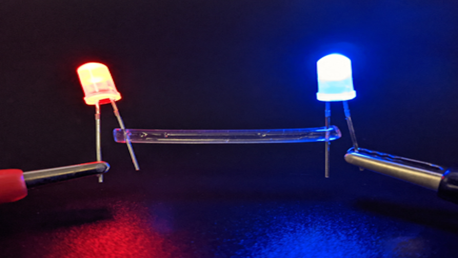
Could Liquid Wires Revolutionize How Doctors Pace the Heart?

A novel hydrogel electrode may improve the effectiveness of electrotherapy.
Cardiac arrhythmias, such as ventricular fibrillation, are the leading cause of sudden cardiac death and are one of the chief causes of morbidity and mortality in the developed world. Arrhythmias can occur when the conduction of electrical signals through the heart tissue is disrupted. This can result from scar tissue, which often forms after a heart attack.
Arrhythmias are commonly treated with cardiac electrotherapies to restore normal conduction. Common electrotherapies, including pace-making, defibrillation, and cardiac resynchronization therapy, involve the delivery of electrical impulses to the heart tissue through lead electrodes, which are surgically inserted in and around the heart.
To maximize the effectiveness of electrotherapies, cardiologists place lead electrodes in areas where they will affect as much heart tissue as possible. When an electrical stimulus induces a group of cardiac cells to contract, those cells are considered “captured” or “recruited” by the stimulus. The more tissue captured, the greater the influence of the electrotherapy and the less damaging the effects of the arrhythmia.
A New Solution Using Injectable Hydrogel Electrodes
Some complex arrhythmias are treated by placing lead electrodes into cardiac veins, which is often the safest way to access the left side of the heart. However, lead electrodes are relatively large compared with these veins, which become even smaller further into the heart. Therefore, leads are placed only in the largest cardiac veins near the heart’s outer surface.
Because the walls of the left ventricle are very thick, lead electrodes placed near the heart’s surface are not able to capture much tissue, limiting the effectiveness of electrotherapies.
Reports have indicated that as many as one-third of patients treated with electrotherapies involving lead placement in the left ventricular wall do not respond well to the treatment.
Thus, a new type of electrode is needed that is capable of traversing more deeply into the cardiac veins than are current lead electrodes.
Researchers led by Dr. Elizabeth Cosgriff-Hernandez at The University of Texas at Austin have partnered with engineers at the Texas Heart Institute (THI) to investigate a novel solution to this problem using injectable hydrogel electrodes. Simply put, hydrogels are polymer networks that swell, with soft-tissue-like properties similar to the heart tissue.
A hydrogel solution injected into a cardiac vein can flow deep into the smallest vessel branches before solidifying into the conductive hydrogel electrode. In this way, the hydrogel electrode can reach much farther into the heart’s deepest and smallest venous branches compared to standard pacemaker leads, resulting in increased capture potential to improve the effectiveness of electrotherapies.
Biomedical engineers from Dr. Cosgriff-Hernandez’s Lab have developed a new type of conductive hydrogel for this purpose. New chemistries were identified that combine conductivity with flexibility, durability, and biocompatibility.
THI Researchers Assess the Performance of Hydrogel Electrodes
To test the hydrogel concept in vivo, researchers in the Electrophysiology Clinical Research and Innovations (EPCRI) Lab at THI headed by Dr. Mehdi Razavi performed experiments in a living heart. Using different types of electrodes, they compared the heart’s response to the delivery of electrical stimuli.
To start, they placed a metal electrode on the surface of the heart (ie, the epicardium) to mimic a standard lead electrode. Electrical stimuli were delivered from the metal electrode, which induced the heart to beat in rhythm with the signals.
Next, the liquid precursor solutions were injected into a large vein. From that point, the precursor solutions traversed through the smaller venous branches deep into the heart, where they reacted to form the conductive hydrogel electrode.
Electrical stimuli were then applied through this hydrogel, which also induced rhythmic beating of the heart.
To assess the relative effectiveness of pacing from the epicardium with a metal electrode or an injected hydrogel electrode, the researchers examined electrograms, which are the electrical signals from the heart as it beats, after using various methods of stimulus delivery.
The electrograms were recorded by the sensing electrodes when the heart was beating normally without external stimuli.
When the electrograms were compared, an exciting pattern was observed. Although each method of electrical stimuli delivery induced the heart to beat, the electrograms resulting from stimuli delivered via the injected hydrogel matched those resulting from natural heartbeats.
On the other hand, the electrograms resulting from epicardial pacing did not match those resulting from natural heartbeats; in fact, they often seemed to be inverted.
This finding implied that epicardial stimuli result in conduction patterns that are very different from natural conduction, whereas stimuli delivered from the hydrogel tended to mimic native conduction.
Why Hydrogel Electrodes May Improve Electrotherapy
To the researchers, it was not surprising that stimuli delivered from a source such as a simple metal electrode produced electrograms indicative of non-physiologic conduction. Patients who have a pacemaker with a single-lead electrode placed in the right ventricle have long been known to be prone to heart failure. The chief culprit is the non-physiologic conduction that results from this kind of pacing. According to the researchers, the far more interesting revelation was that pacing from the injected hydrogel seemed to result in conduction patterns that closely resembled healthy physiologic conduction. This finding may have significant implications for future electrotherapies.
To understand the potential mechanism underlying this revelation, consider a healthy heart, in which electrical signal conduction from cell to cell does not happen like a game of telephone. Instead, conduction is facilitated by a branching network of nerves that coordinate to optimize the overall contraction of the heart. Electrical impulses originate at a special bundle of nerves called the sinoatrial (SA) node, also known as the natural pacemaker of the heart. From the SA node, signals are conducted faster than in normal heart cells, providing a pathway that is preferentially followed by a natural electrical impulse. As the impulse travels along this pathway, nearby cells are activated in a coordinated cascade that results in a natural, healthy heartbeat.
When an electrical impulse is not generated by the SA node but rather by an external source, such as a lead electrode placed in an epicardial vein, the impulse is not connected to the pathway of fast-conducting nerves that facilitates healthy electrical conduction.
Because these nerve pathways are primarily positioned in the middle of the heart tissue in a region known as the mid-myocardium, electrodes near the heart’s surface cannot mimic the conduction patterns associated with healthy heartbeats. Rather, the impulse propagates blindly through the heart tissue, inducing the heart to beat in an uncoordinated (and potentially unhealthy) manner.
A key advantage of the hydrogel electrode system is that, because of its injectability, it can penetrate deeper into the heart tissue than is possible with current lead electrodes. Before the liquid precursors react to solidify into the final hydrogel, they can flow into even the smallest of vessels deep in the heart tissue, including the mid-myocardial region where the nerve pathway is located.
The branches of nerve fibers that service the ventricles (ie, the larger, thicker heart chambers responsible for providing blood to the lungs and body) are called Purkinje fibers. When an electrode located near Purkinje fibers is stimulated, the native conduction pathway may be accessed, resulting in heartbeats that bear a close resemblance to healthy, natural heart beats.
According to researchers in the EPCRI Lab at THI, this mechanism could be responsible for the remarkable similarities between electrograms recorded from natural heartbeats and from heartbeats induced by delivering stimuli through hydrogel electrodes.
Potential Indications for Hydrogel Electrodes
The early results of experiments with the conductive hydrogel electrode have profound implications in the field of electrophysiology. They suggest that not only can pacing therapy be safely and effectively delivered through the novel hydrogel material, but the hydrogel can penetrate deep into the small vessels of the mid-myocardium, enabling far greater capture potential than is currently possible with modern electrodes.
Further, by capturing the native conduction pathway directly, this electrode can induce a more physiologic heartbeat than that generated from standard electrodes. Thus, using a hydrogel electrode may alleviate some of the adverse effects of long-term pacing in the ventricles.
There are numerous potential applications of the novel hydrogel electrode that warrant further investigation. Patients with ischemic heart attacks often develop scar tissue around the blocked coronary artery where the blood supply to the heart tissue was cut off.
These scarred regions disrupt impulse conduction and are common sites for cardiac arrhythmias to develop. By placing a hydrogel electrode in a vein that crosses one of these scarred regions, it may be possible to bridge zones of healthy tissue, bypassing the scarred region and restoring healthy conduction.
Other patients who undergo advanced pacing therapies involving left ventricular leads may benefit from replacing these leads with a hydrogel electrode, which can capture more heart tissue in a physiologic fashion.
Treatments such as cardiac resynchronization therapy with limited efficacy often attributed to poor left ventricular lead function may show improved success.
Furthermore, the hydrogel electrode may have implications for defibrillation. The complex emergent circuits responsible for arrhythmias such as ventricular fibrillation are often anchored in the mid-myocardial regions, which are made accessible via the hydrogel.
Electrotherapy with the hydrogel may terminate arrhythmias with far less energy than current defibrillators. Because shocks delivered from standard implanted defibrillators are very painful and occur without warning, electrotherapy with a hydrogel electrode would be of significant interest to patients and doctors alike.
Future Directions
Much work remains to be done to translate these exciting early results into a therapy that can be used reliably in human patients. First, the chemistry of the hydrogel must be further refined for chronic application. Although early indications of the hydrogel are favorable in terms of biocompatibility, stability, and mechanical robustness, long-term trials in animal models are needed to show that these qualities can be maintained throughout a patient’s lifetime.
Additionally, a reliable catheter-based system for injecting the precursor solutions is currently under development by engineers in the EPCRI Lab at THI so that the hydrogel can be delivered via a minimally invasive procedure. Because the in situ hydrogel method relies on the in-vessel reaction between two precursor solutions, a reliable system is needed for delivering these precursors to the target vessel without allowing them to interact before injection.
Further studies will be underway in the near future to investigate and clarify these concerns. The teams led by Drs. Cosgriff-Hernandez and Razavi believe that the hydrogel technology may provide a platform for a new generation of cardiac electrotherapies.


