Catheter Ablation
Catheter ablation is a procedure where energy is used to treat the site of an arrhythmia. Several different forms of energy can be used for this purpose, but the most common type is radiofrequency energy. Radiofrequency energy is a type of energy that uses radio waves to produce heat that destroys the tiny area of heart tissue that is causing your arrhythmia. When the tissue is destroyed, your heart can return to its regular rhythm. This procedure is also called radiofrequency ablation.
Any irregularity in your heart’s natural rhythm is called an arrhythmia. Almost everyone’s heart skips beats, and these mild palpitations are usually harmless. But there are about 4 million Americans who have recurrent arrhythmias, and these people usually need treatment for their condition.
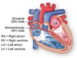
Electrical impulses from the heart muscle cause your heart to beat (contract). This electrical signal begins in the sinoatrial (SA) node, located at the top of the heart’s upper-right chamber (the right atrium). The SA node is sometimes called the heart’s “natural pacemaker.”
The SA node sends electrical impulses at a certain rate, but your heart rate may still be altered by physical demands, stress, or other factors. Sometimes, the SA node does not work properly, causing the heart to beat too fast, too slow, or irregularly. In other cases, the heart’s electrical pathways are blocked, which can also cause an irregular heart rhythm.When an electrical impulse is released from the SA node, it causes the upper chambers of the heart (the atria) to contract. The signal then passes through the atrioventricular (AV) node. The AV node checks the signal and sends it through the muscle fibers of the lower chambers (the ventricles), causing them to contract.
For most people with arrhythmias, medicines work quite well to control the abnormal heart rhythm. But when medicines don’t work, doctors may suggest catheter ablation.
What is catheter ablation?
Catheter ablation is a procedure where energy is used to treat the site of an arrhythmia. Several different forms of energy can be used for this purpose, but the most common type is radiofrequency energy. Radiofrequency energy is a type of energy that uses radio waves to produce heat that destroys the tiny area of heart tissue that is causing your arrhythmia. When the tissue is destroyed, your heart can return to its regular rhythm. This procedure is also called radiofrequency ablation.
Catheter ablation is used to treat a wide variety of cardiac arrhythmias. Some of them include:
- Supraventricular tachycardia (SVT) is a rapid, regular heart rate where the heart beats anywhere from 150-250 times per minute in the atria. Another name for SVT is paroxysmal supraventricular tachycardia (PSVT). The word “paroxysmal” means occasionally or from time to time.
- Atrial flutter happens when the atria beat very fast, causing the ventricles to beat inefficiently as well.
- Atrial fibrillation is a fast, irregular and chaotic rhythm involving the atrial chambers. It results in loss of pumping contribution by the atria and rapid irregular beating of ventricles. It is a main cause of stroke, especially among elderly people.
- Ventricular tachycardia is rapid heartbeat arising from the lower chambers of the heart.
What can I expect during catheter ablation?
The procedure is performed in the cardiac catheterization angiography suite (also called the cath lab) or the electrophysiology lab. You may not eat or drink anything after midnight the night before the procedure. If you have diabetes, you should talk to your doctor about your food and insulin intake, because not eating can affect your blood sugar levels.
Talk to your doctor about any medicines (prescription, over-the-counter, or supplements) that you are taking. This is especially important if you are taking blood-thinning medicines (anticoagulants) or antiplatelet therapy.
Once you are in the cath lab, you will see television monitors, heart monitors, and blood pressure machines. You will lie on an examination table, which is usually near an x-ray camera. Small metal disks called electrodes will be placed on your chest. These electrodes hook up to an electrocardiogram machine which monitors your heart rhythm during the procedure. To prevent infection, you will be shaved and cleansed around the area where the catheter will be inserted.
A very small needle will be put in the vein of your forearm. This is called an intravenous line or IV. The IV will be used to give you a medicine to relax you, and you may even fall asleep. Otherwise, you will be awake for the procedure.
You will be given an anesthetic medicine to numb the area around where the catheter will be inserted. This needle may hurt a bit, much like at the dentist after being given Novocain. You should not feel pain during any part of the procedure, but if you do, ask the doctors for more medicine.
After gaining entrance into a blood vessel in your groin, arm, or neck, doctors insert several long, thin tubes with wires, called electrode catheters, through a sheath and feed these tubes into your heart. They use a video monitor (like a TV screen) to see the process. You will not feel the catheter passing through the blood vessel because it lacks nerve endings on the inside lining.
To locate the abnormal tissue causing arrhythmia, doctors send a small electrical impulse through the electrode catheter to activate your arrhythmia. Other catheters are used to record the heart’s electrical signals and locate the abnormal tissue. Doctors will then place a catheter at the exact site of the abnormal cells in your heart. The radiofrequency energy is sent through this catheter to cauterize the tissue causing your arrhythmia. You may feel a mild burning sensation when the radiofrequency energy is applied to the heart tissue.
After the catheter is removed, firm pressure will be applied to the site in your groin to stop any bleeding. You will also be bandaged. To avoid bleeding at the catheter insertion site, you will need to lie very still for several hours, either in the recovery area or in your hospital room.
Catheter ablation usually takes 2 to 4 hours. If you have several areas of abnormal tissue, then the procedure may take longer. Depending on how you are feeling after the procedure, you may either go home the same day or you may have to stay overnight in the hospital.
What happens after the procedure?
After you leave the hospital, your doctor will give you specific instructions about drinking plenty of fluids, driving, and bathing. Most people can return to their normal activities the day after they leave the hospital. You should avoid heavy physical activity for 2-3 days. Ask your doctor when you can return to strenuous exercise.
You may feel fatigue or chest discomfort for about 2 days after the procedure. You should tell your doctor if this lasts longer than 2 days or the pain is severe. You may also experience palpitations after the procedure. These abnormal heartbeats will go away as your heart heals after the procedure.


